| Parasite | Didymocystis wedli |
|---|---|
| Taxonomy | Plathyhelminthes, Trematoda, Didymozoidae |
| Host | Northern bluefin tuna (Thunnus thynnus) |
| Infection site | Gill filament |
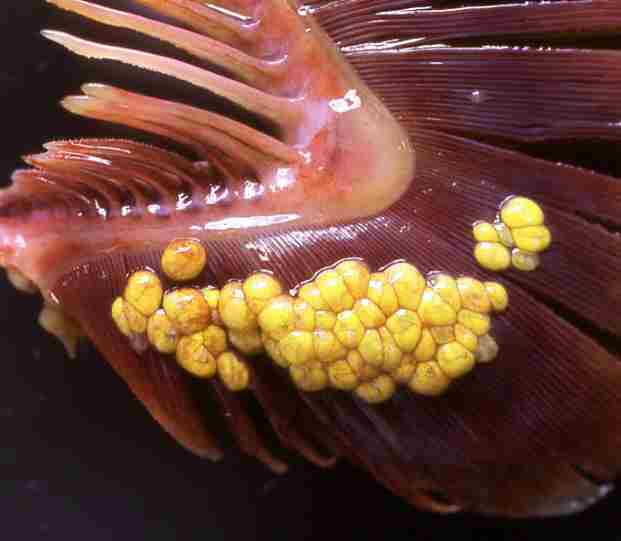
| Clinical sign | Several dozens of yellowish cysts (ca. 2.0×2.5 mm) are observed in the gill filaments (Fig. 1). |
| Parasitology | A cyst includes one male and one female. They are placed side-by-side as if they hold each other. (Fig. 2). The body is 2.5 mm long. The parasite has a number of kidney bean-shaped eggs (ca. 20 mm × 12.5 mm) (Fig. 3). The life cycle is unknown. |
| Pathology | Intensity of infection is not correlated with the growth of fish and BMI, suggesting that pathogenic effects of this parasite is low (Momoyama and Kobayashi, 2004). |
| Health hazard | Since this parasite is not infectious to human, it is harmless in food hygiene. |
| Diagnosis | Check the cysts and morphology of the parasite in the gill. |
| Other information | As growing interests on aquaculture of Northern bluefin tuna using the wild seedlings, this parasite has been frequently found. Some other didymozooidae trematodes were also reported in Northern bluefin tuna, Didymocystis semiglobularis in the epidermis of the gill arch (Fig. 4); Didymocystis sloeiformis in the bone tissue of the gill arch (Fig. 5); Kollikeria reniformis at the surface of the gill raker (Fig. 6); Coeliotrema thynnus in the pyloric caecum; Wedlia orientalis in the gastric submucosa. |
| References | Momoyama, K. and T. Kobayashi (2004): Didymozoid
parasites found in the blue fin tuna,
Thunnus thynnus, caught in the coastal waters along Yamaguchi Prefecture in the Japan
Sea.
Bull. Yamaguchi Pref. Fish. Res. Ctr., 2, 125-132. |

(Photos by K. Momoyama)
Fig. 7. K. reniformis on the gill raker
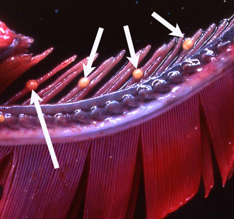
Fig. 5. D. semiglobularis
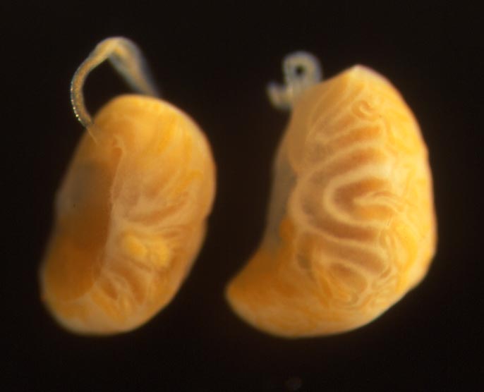
Fig. 1. D. wedli parasitizing the gill filament of Norhtern bluefin tuna
Fig. 2. A pair of D. wedli (arrows) released from the transparent membrane (*) of a cyst.
Fig. 6. D. soleiformis in the bone tissue of gill arch
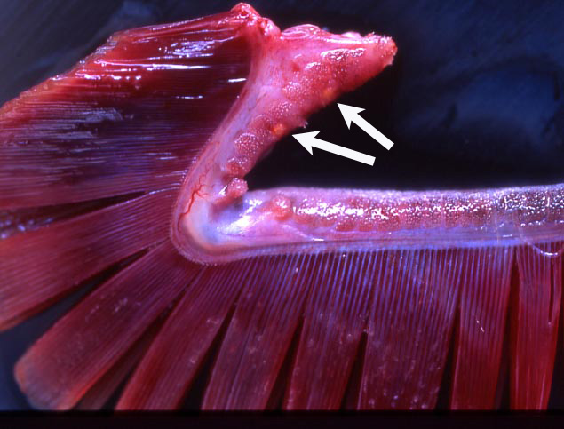
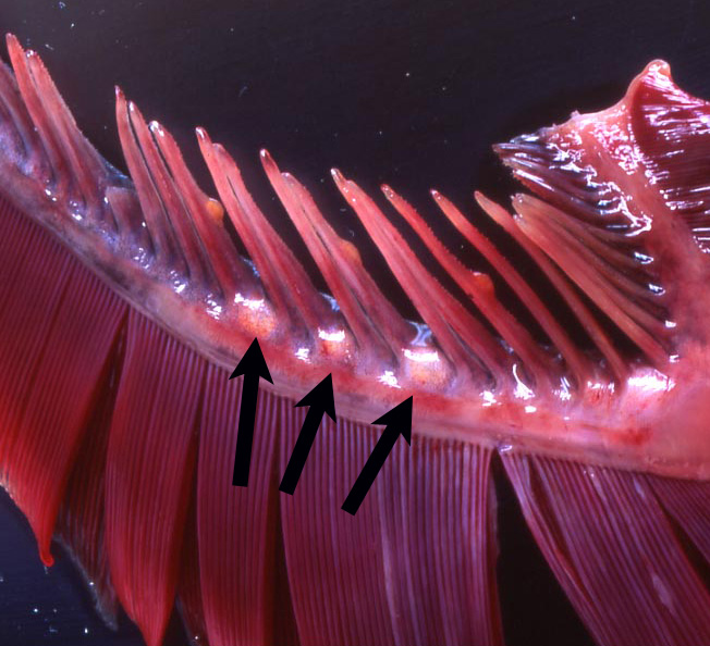
Fig. 3. Eggs of D. wedli
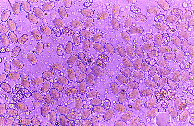
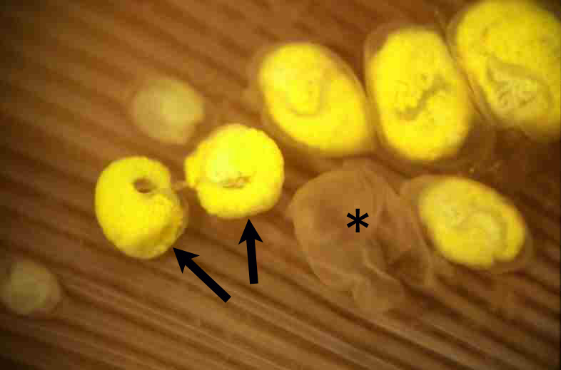
Fig. 4. D. semiglobularis (arrows) on the gill arch.